


Fetal ultrasound…safe way to check your baby’s health
Sunday, June 30, 2019
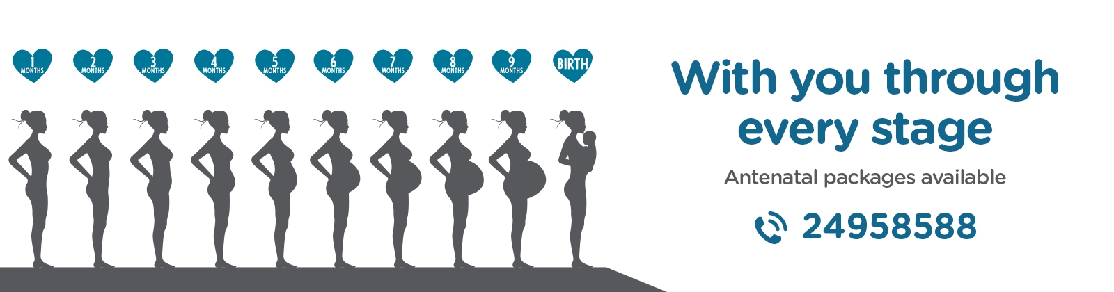
Fetal ultrasound is a test used during pregnancy. It creates an image of the baby in the mother’s womb (uterus). It provides a safe way to evaluate the health of an unborn baby. During a fetal ultrasound, the baby’s heart, head, and spine are evaluated, along with other parts of the baby. The test may be done either on the mother’s abdomen (transabdominal) or in the vagina (transvaginal).
Why might I need a fetal ultrasound?

Fetal ultrasound is a routine part of prenatal care. This is because it’s a low-risk procedure that gives important information. A routine prenatal ultrasound can check for defects or other problems in the fetus. As the pregnancy progresses, ultrasound becomes more and more accurate for determining the viability of a pregnancy. If an ultrasound in the second or third trimester shows that a baby has no heartbeat, this is considered conclusive for diagnosing a missed miscarriage.
The Following can be examined:
- Head and brain: The chambers within the brain (ventricles), distance between parietal bones of the fetal head (biparietal diameter), and skin thickness at the back of the head (nuchal area) is evaluated for defects.
- Heart: The chambers and valves of the heart are evaluated and defects may be identified.
- Abdomen and stomach: The size, location, and arrangement of stomach and diaphragm are checked.
- Urinary bladder: The size and presence of the bladder is evaluated.
- Spine: Defects may be identified if present.
- Umbilical cord: Three blood vessels should be attached at the front of the abdomen.
- Kidneys: Two kidneys should be present on either side of the mid-spine.
- Other fetal structures: Limbs and other parts may also be scanned and evaluated
A fetal ultrasound can also show:
- If a woman is pregnant with multiple babies
- The gestational age of a baby
- Where to place the needle during removal of amniotic fluid (amniocentesis)
There are several types of fetal ultrasound:
- Standard ultrasound: The test uses sound waves to create two-dimensional images on a computer screen.
- Doppler ultrasound: This test shows the movement of blood through the umbilical cord, in the baby’s heart, or between the baby and the placenta.
- 3 & 4 D ultrasound: This test shows a lifelike image of an unborn baby.
- Fetal echocardiography. This exam provides a detailed picture of a baby’s heart. It might be used to confirm or rule out a congenital heart defect.
- Fetal Echocardiography: This exam provides a detailed picture of a baby’s heart. It might be used to confirm or rule out a congenital heart defect.